Home
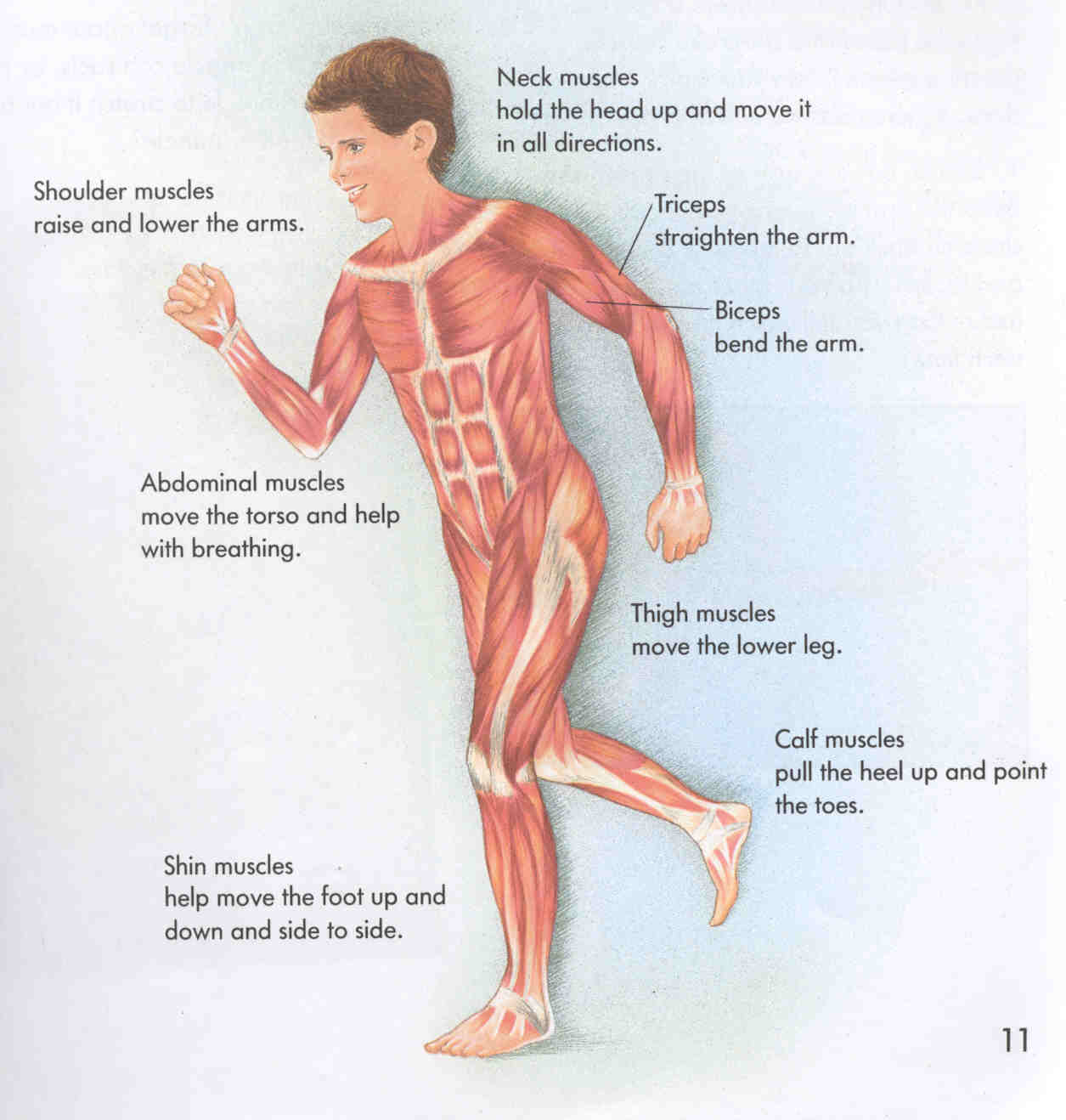
/ Muscle Diagram Labeled - Labeled Muscles Of Lower Leg Human Muscle Anatomy Human Body Muscles Human Muscular System, These muscles can be grouped based upon their location and function.
Muscle Diagram Labeled - Labeled Muscles Of Lower Leg Human Muscle Anatomy Human Body Muscles Human Muscular System, These muscles can be grouped based upon their location and function.
Muscle Diagram Labeled - Labeled Muscles Of Lower Leg Human Muscle Anatomy Human Body Muscles Human Muscular System, These muscles can be grouped based upon their location and function.. Spend some time revising this diagram by connecting the name and location of each structure with what you've just learned in the video. There are around 650 skeletal muscles within the typical human body. Find a great range of the diagram of human body and anatomy diagrams in the following pictures. Muscle diagram blank each of. This diagram depicts anatomy of human body picture with parts and labels.
Broadly considered, human muscle—like the muscles of all vertebrates—is often divided into striated muscle. For more anatomy content please follow us and visit our website: Muscle diagrams are a great way to get an overview of all of the muscles within a body region. When you are taking anatomy and physiology you will be required to identify major muscles in the human body. You go to the gym to train your abs.
Labeled Diagram Of Muscular System Biological Science Picture Directory Pulpbits Net from pulpbits.net The many muscles of the hip provide movement, strength, and stability to the hip joint and the bones of the hip and thigh. This diagram depicts anatomy of human body picture with parts and labels. The axial skeleton runs along the body's midline axis and is made up of 80 bones in the following regions: There are 20 muscle pairs, one on each side of the body. Thigh muscle anatomy knee muscles anatomy shoulder muscle anatomy skeletal muscle anatomy human muscle anatomy human anatomy and physiology body anatomy leg muscles diagram muscle diagram. Skeletal muscles attach to and move bones by contracting and relaxing in response to voluntary messages from the nervous system. Once you're feeling confident, it's time to test yourself. Within this group of back muscles you will find the latissimus dorsi, the trapezius, levator scapulae and the rhomboids.
I mean, the abs are the muscle.
You go to the gym to train your abs. There are around 650 skeletal muscles within the typical human body. Activity 4.6 labeled muscle diagram. A muscle of the leg originating on the lateral condyle of the tibia and the interosseus membrane between the tibia and the fibula and inserted in the first cunieform and first metatarsal bones. Discover the muscle anatomy of every muscle group in the human body. Major posterior muscles | anatomy. Muscle fibers are organized into bundles supplied by blood vessels and innervated by motor neurons. Muscle diagram blank each of. When you are taking anatomy and physiology you will be required to identify major muscles in the human body. There are 20 muscle pairs, one on each side of the body. 12 photos of the muscles labeled front and back. Broadly considered, human muscle—like the muscles of all vertebrates—is often divided into striated muscle, smooth muscle, and cardiac muscle. The human body is one complex network, universally accepted as the most intriguing construct.
Skeletal muscles attach to and move bones by contracting and relaxing in response to voluntary messages from the nervous system. Take a look at the leg muscles diagram below, where you see each muscle clearly labeled. Once you're feeling confident, it's time to test yourself. Activity 4.6 labeled muscle diagram. The interactive muscle anatomy diagram shown below outlines the major superficial (i.e.
Human Arm Muscles Diagram Anatomical Models Ball State University Digital Media Repository from dmr.bsu.edu It dorsi flexes and inverts the foot, supports the arch, and is controlled by the deep peroneal. Home › training design › anatomy and physiology › muscle charts of the human body. Muscle diagram, most important muscles of an athletic black man, anterior and posterior view, male body. Skeletal muscle tissue is composed of long cells called muscle fibers that have a striated appearance. There are 20 muscle pairs, one on each side of the body. This might sound like a strange question, right? This diagram depicts muscle labeled diagram.human anatomy diagrams show internal organs, cells, systems, conditions, symptoms and sickness information and/or tips for healthy living. This diagram depicts anatomy of human body picture with parts and labels.
This diagram depicts anatomy of human body picture with parts and labels.
Broadly considered, human muscle—like the muscles of all vertebrates—is often divided into striated muscle, smooth muscle, and cardiac muscle. You can click the links in the image, or the links below the image to find out more information on any muscle group. Use the location, shape and surrounding structures to help you memorize each muscle. I mean, the abs are the muscle. Muscle fibers are organized into bundles supplied by blood vessels and innervated by motor neurons. How many muscles are in the back? Within this group of back muscles you will find the latissimus dorsi, the trapezius, levator scapulae and the rhomboids. Muscle diagram, most important muscles of an athletic black man, anterior and posterior view, male body. We think this is the. Activity 4.6 labeled muscle diagram. When you are taking anatomy and physiology you will be required to identify major muscles in the human body. You go to the gym to train your abs. It dorsi flexes and inverts the foot, supports the arch, and is controlled by the deep peroneal.
Spend some time revising this diagram by connecting the name and location of each structure with what you've just learned in the video. The lower part of the trapezius ascends and depresses the scapula, while the transverse or middle region of the trapezius is what retracts the. For your reference value these charts show the major superficial and deep muscles of the human body. Within this group of back muscles you will find the latissimus dorsi, the trapezius, levator scapulae and the rhomboids. The axial skeleton and the appendicular skeleton.
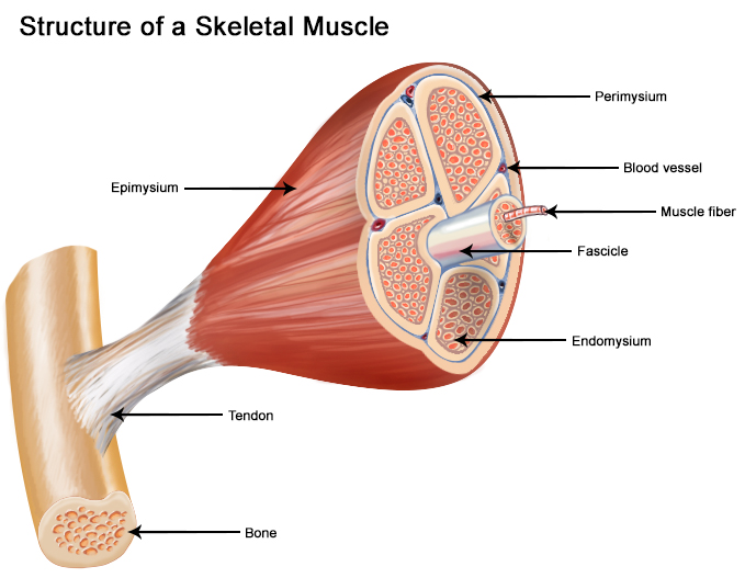
Seer Training Structure Of Skeletal Muscle from training.seer.cancer.gov There are three parts to the trapezius. Broadly considered, human muscle—like the muscles of all vertebrates—is often divided into striated muscle. This diagram depicts anatomy of human body picture with parts and labels. This quiz requires labeling, so it will test your knowledge on how to identify these muscles (latissimus dorsi, trapezius, deltoid, biceps brachii, triceps brachii, brachioradialis, pectoralis major, serratus anterior, rectus abdominis, etc.). The back has a total of 40 muscles. Likewise, there are muscles in other parts of the body that help support and move the spine. Home › training design › anatomy and physiology › muscle charts of the human body. You go to the gym to train your abs.
When you are taking anatomy and physiology you will be required to identify major muscles in the human body.
There are 20 muscle pairs, one on each side of the body. When you think of abs, what muscle do you typically think of? 12 photos of the muscles labeled front and back. It dorsi flexes and inverts the foot, supports the arch, and is controlled by the deep peroneal. Muscle fibers are organized into bundles supplied by blood vessels and innervated by motor neurons. Discover the muscle anatomy of every muscle group in the human body. The axial skeleton and the appendicular skeleton. I mean, the abs are the muscle. The majority of muscles in the leg are considered long muscles, in that they stretch great distances. The four groups are the anterior group, the posterior group, adductor group, and finally the abductor group. The many muscles of the hip provide movement, strength, and stability to the hip joint and the bones of the hip and thigh. Below you'll see diagrams along with the names of the back muscles that may be the cause of your pain. The lower part of the trapezius ascends and depresses the scapula, while the transverse or middle region of the trapezius is what retracts the.